
Paper Published from Lab Visiting Program
Novel mutations in malonyl-CoA-acyl carrier protein transacylase (MCAT) provoke autosomal recessive optic neuropathy.
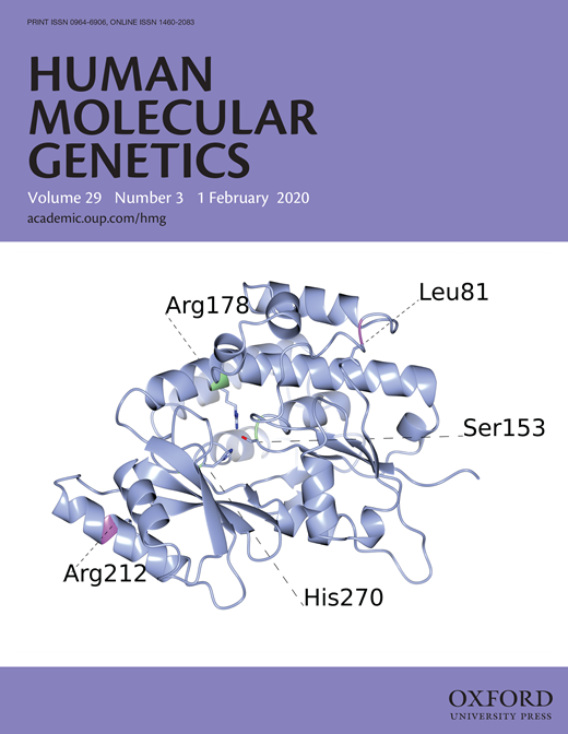 Cover of Journal: MCAT Protein Structure
Hum Mol Genet. 2020 Feb 1;29(3):444-458. doi: 10.1093/hmg/ddz311.
Li H(1)(2), Yuan S(1)(2), Minegishi Y(1), Suga A(1), Yoshitake K(1), Sheng X(2),
Ye J(3), Smith S(4), Bunkoczi G(5), Yamamoto M(1), Iwata T(1).
(1)Division of Molecular and Cellular Biology, National Institute of Sensory Organs, National Hospital Organization Tokyo Medical Center, 2-5-1, Higashigaoka, Meguro-ku, Tokyo, 152-8902, Japan.
(2)Ningxia Clinical Research Center of Blinding Eye Disease, Ningxia Eye Hospital, People Hospital of Ningxia Hui Autonomous Region, No. 936, Huang He East Road,Yinchuan, 750001, China.
(3)Pennington Biomedical Research Center, Louisiana State University Systems, 6400, Perkin Road, Baton Rouge, LA, 70808, USA.
(4)Children's Hospital Oakland Research Institute, 5700, Martin Luther King Jr. Way, Oakland, CA, 94609, USA.
(5)Astex Pharmaceuticals, 436, Cambridge Science Park, Cambridge, CB4 0QA, UK.
Inherited optic neuropathies are rare eye diseases of optic nerve dysfunction that present in various genetic forms. Previously, mutation in three genes encoding mitochondrial proteins has been implicated in autosomal recessive forms of optic atrophy that involve progressive degeneration of optic nerve and retinal ganglion cells (RGC). Using whole exome analysis, a novel double homozygous mutation p.L81R and pR212W in malonyl CoA-acyl carrier protein transacylase (MCAT), a mitochondrial protein involved in fatty acid biosynthesis, has now been identified as responsible for an autosomal recessive optic neuropathy from a Chinese consanguineous family. MCAT is expressed in RGC that are rich in mitochondria. The disease variants lead to structurally unstable MCAT protein with significantly reduced intracellular expression. RGC-specific knockdown of Mcat in mice, lead to an attenuated retinal neurofiber layer, that resembles the phenotype of optic neuropathy. These results indicated that MCAT plays an essential role in mitochondrial function and maintenance of RGC axons, while novel MCAT p.L81R and p.R212W mutations can lead to optic neuropathy.
Mutation in the zebrafish cct2 gene leads to abnormalities of cell cycle and cell death in the Retina: A model of CCT2-related Leber Congenital Amaurosis
Invest Ophthalmol Vis Sci. 2018 Feb 1;59(2):995-1004. doi: 10.1167/iovs.17-22919.
Minegishi Y(1), Nakaya N(1), Tomarev SI(1).
(1)Section of Retinal Ganglion Cell Biology, Laboratory of Retinal Cell and Molecular Biology, National Eye Institute, National Institutes of Health, Bethesda, Maryland, United States.
The compound heterozygous mutations in the β subunit of chaperonin containing TCP-1 (CCT), encoded by CCT2, lead to the Leber congenital amaurosis (LCA). In this study, a cct2 mutant line of zebrafish was established to investigate the role of CCT2 mutations in LCA in vertebrates. A cct2 mutant zebrafish line was produced using the CRISPR-Cas9 system. Changes in the eyes of developing wild-type and mutant larvae were monitored using microscopy, immunostaining, TUNEL, and EdU assays. Phenotypic rescue of mutant phenotype was investigated by injection of CCT2 RNA into zebrafish embryos. The cct2 mutation (L394H-7del) led to the synthesis of a mutated cctβ protein with the L394H replacement and deletion of 7 amino acid residues (positions 395-401). The homozygous cct2-L394H-7del mutant exhibited a small eye phenotype at 2 days post fertilization (dpf) and was embryonically lethal after 5 dpf. In homozygous cct2-L394H-7del mutants, the retinal ganglion cell differentiation was attenuated, retinal cell cycle was affected, and the neural retinal cell death was significantly increased at 2 dpf compared with wild-type. Injection of RNA encoding wild-type human CCTβ rescued the small eye henotype, reduced retinal cell death, and restored the levels of CCTβ protein and the major client protein Gβ1 that were significantly reduced in the homozygous cct2-L394H-7del mutant compared with wild-type. These results indicate that cct2 plays an essential role in retinal development by regulating the cell cycle. The retinal pathology observed in the homozygous cct2-L394H-7del mutants resembles the retinal pathology of human LCA patients.
CCT2 Mutations Evoke Leber Congenital Amaurosis due to Chaperone Complex Instability
Sci Rep. 2016 Sep 20;6:33742. doi: 10.1038/srep33742.
Minegishi Y(1), Sheng X(2), Yoshitake K(3), Sergeev Y(4), Iejima D(1), Shibagaki Y(5), Monma N(3), Ikeo K(3), Furuno M(6), Zhuang W(2), Liu Y(2), Rong W(2), Hattori S(5), Iwata T(1).
(1)Division of Molecular and Cellular Biology, National Institute of Sensory Organs, National Hospital Organization Tokyo Medical Center, Tokyo, Japan. (2)Ningxia Eye Hospital, Ningxia People's Hospital, Ningxia, China. (3)Laboratory of DNA Data Analysis, National Institute of Genetics, Shizuoka, Japan. (4)National Eye Institute, National Institutes of Health, Bethesda, MD, USA. (5)Division of Biochemistry, School of Pharmaceutical Science, Kitasato University, Tokyo, Japan. (6)RIKEN Center for Life Science Technologies, Division of Genomic Technologies, Life Science Accelerator Technology Group, Transcriptome Technology Team, Yokohama, Japan.
Leber congenital amaurosis (LCA) is a hereditary early-onset retinal dystrophy that is accompanied by severe macular degeneration. In this study, novel compound heterozygous mutations were identified as LCA-causative in chaperonin-containing TCP-1, subunit 2 (CCT2), a gene that encodes the molecular chaperone protein, CCTβ. The zebrafish mutants of CCTβ are known to exhibit the eye phenotype while its mutation and association with human disease have been unknown. The CCT proteins (CCT α-θ) forms ring complex for its chaperon function. The LCA mutants of CCTβ, T400P and R516H, are biochemically instable and the affinity for the adjacent subunit, CCTγ, was affected distinctly in both mutants. The patient-derived induced pluripotent stem cells (iPSCs), carrying these CCTβ mutants, were less proliferative than the control iPSCs. Decreased proliferation under Cct2 knockdown in 661W cells was significantly rescued by wild-type CCTβ expression. However, the expression of T400P and R516H didn't exhibit the significant effect. In mouse retina, both CCTβ and CCTγ are expressed in the retinal ganglion cells and connecting cilium of photoreceptor cells. The Cct2 knockdown decreased its major client protein, transducing β1 (Gβ1). Here we report the novel LCA mutations in CCTβ and the impact of chaperon disability by these mutations in cellular biology.